High Performance Liquid Chromatography: An Alternative Method for Identifying Bloodstain Origins in Wildlife Forensics
Introduction
The high frequency of occurrence of bloodstains at crime scenes has resulted in an increased need for sensitive identification methods. In the case of wildlife forensic evidence, this translates to specific techniques capable of identifying the source species (animal) of a bloodstain. Standard techniques include immunodiffusion, immunoelectrophoresis, and isoelectric focusing. Each of these techniques has certain limitations.
Order custom essay High Performance Liquid Chromatography: An Alternative Method for Identifying Bloodstain Origins in Wildlife Forensics with free plagiarism report
 450+ experts on 30 subjects
 Starting from 3 hours delivery
The application of another analytical technique, High Performance Liquid Chromatography (HPLC), apparently overcomes many of these inherent limitations, to provide a rapid, efficient and sensitive method for the determination of origin of bloodstains. Article Summary : Blood evidence is regularly examined in the forensic wildlife community.
As such, sensitive and accurate techniques must be employed. This article examines the techniques currently being used and their limitations, and offers an alternative analytical technique, High Performance Liquid Chromatography. Use of this technique avoids the necessity for specific antisera, required for previously employed immunological methods. In this report, the analysis of 275 animal samples (262 individual samples and 13 mixtures) by HPLC is considered, and the benefits explained. The hemoglobin of 22 discrete families comprising 38 genera and approximately 50 different species was collected, from both wet and dry samples.
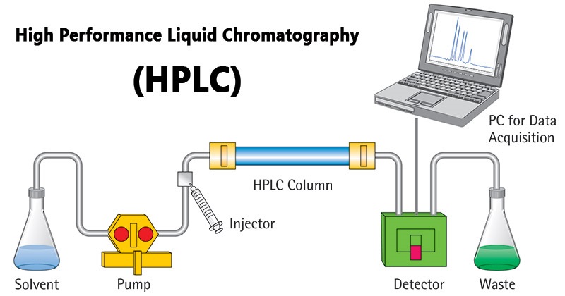
A Hewlett Packard 1090 Series II HPLC with a diode-array detector set to detect the various components of hemoglobin was used. Solvents were applied using the gradient method, enabling high resolution separation of the specific components. Analysis by HPLC of these different animal hemoglobin samples resulted in species specific retention times for the 1-globin, 1-globin and heme components of the hemoglobin. "The unique chromatographic retention times produced by the globin chain signals from species to species serve as the profiles to which an unknown species is compared and identified." The chromatographic results obtained using HPLC were reproducible, with the heme signal for all samples appearing at the same retention time (8.22 min + 0.18 min), and samples as small as 1.2 ig of hemoglobin were able to be analysed. Reasonably good determination (quantitation) of hemoglobin quantities was also possible using this method, as was identification of blood mixtures. On a whole, the technique of HPLC provided a rapid, sensitive and reproducible method for the identification and quantitation of animal blood samples. Analytical Methods: 1)
Immunodiffusion Precipitating antibody is often produced during the normal sequence of events in an infection. When rabbits are injected with the blood from another animal, antibodies are formed that react with the invading animal blood to neutralise its presence. These antibodies can be isolated, and used in further analysis, as they will react specifically with antigens of that particular animal species. The process of immunodiffusion takes advantage of the fact that antibodies and antigens will diffuse towards each other on an agar gel-coated plate. Antiserum and the extracted blood evidence are placed in separate holes on the gel. Eventually, antibodies from the antiserum and antigens meet in the gel. If the antibodies for the specific antigens exist in the antiserum, they will bind with that antigen. Agglutination occurs if the antigen is recognized, possible as a result of the antibodies being bivarient.
This agglutination results in a visible line between the two wells to which the antiserum and the recognized antigen were originally added. The major limitation of this technique is the necessity for known antiserum. While there are a number of animal antiserums widely available, they are not obtainable for all wildlife families. 2) Immunoelectrophoresis Immunoelectrophoresis combines the process of serum electrophoresis and an antigen-antibody interaction. Electrophoresis is a separation technique that is based on the mobility of ions in an applied electric field. Positively charged ions migrate towards a negative electrode and negatively-charged ions migrate toward a positive electrode. The ions have different migration rates depending on their total charge, size, and shape, and can therefore be separated. In this case, the blood serum is placed on a barbitone impregnated gel and exposed to an electric current, upon which the various proteins (antigens) migrate to form bands, or zones.
Antisera is then added to a trough in the electrophoretogram, and subsequently diffuses toward the antigen zones. A reaction occurs upon contact, resulting in the precipitation of antigen-antibody complexes. The presence of a precipitin band indicates that the antigen is present for specific antiserum used. Once again, this procedure is limited to analysis with those readily available antisera. 3) Isoelectric Focusing Isoelectric focusing is a well-established technique for the separation of amphoteric molecules such as proteins. Separation is carried out on gels on which a stable pH gradient has been established.
Every ampholytic compound has a specific pH at which it is neutral, called its isoelectric point. The process of isoelectric focusing takes advantage of this fact. The pH gradient is achieved via impregnation of the gel with polyamino-polycarboxylic acids with pls covering the pH range in question. When the electric field is applied, these ampholytes migrate and order themselves across the gel in increasing pl. The strong buffering capacity of these compounds results in the formation of a corresponding pH gradient. When the proteins are applied, they migrate in the applied field and come to rest at the point where the pH is equal to the individual pls. At this point, the compound is converted to its zwitterion form, is neutral, and thus loses its electrophoretic mobility. As proteins are amphoteric they will behave in an identical manner and be focused in a position dictated by their isoelectric point. The high resolution associated with this method is aided by the fact that an amphoteric molecule diffusing away from the position corresponding to its electric point to a region of higher or lower pH will regain a charge and be moved smartly back into line.
As previously stated, high resolution can be achieved using this method, and proteins that differ by as little as 0.01pH units can be adequately resolved. There are, however, a number of limitations of use in forensic applications. Isoelectric focusing has been found to have limited use in differentiation of neutral amino acid substitutions in two otherwise similar proteins and therefore does not distinguish differences in globin molecules completely. Isoelectric focusing is conventionally carried out using either polyacrylamide or agarose as a support medium, and while the support medium is necessary to counteract the excesses of the electric-field-induced convection and diffusion, it does hinder mobility and thus increase the time taken for larger molecules to reach the position of the isoelectric pH. The process is also only applicable if there are stable and detectable species-specific protein markers still present in the material to be analysed. Substantial degradation of the blood evidence may result in the absence of such markers.
Reverse-Phase High Performance Liquid Chromatography
A hemoglobin molecule consists of four polypeptide chains: two alpha chains, each with 141 amino acids and two beta chains, each with 146 amino acids. The protein portion of each of these chains is called "globin". Each globin chain folds into 8 1 helical segments (A-H) which, in turn, fold to form globular tertiary structures. The folded helices form a pocket that holds the working part of each chain, the heme. The heme group is a flat ring molecule containing carbon, nitrogen and hydrogen atoms, with a single Fe2+ ion at the center. Without the iron, the ring is called a porphyrin. In a heme molecule, the iron is held within the flat plane by four nitrogen ligands from the porphyrin ring. The iron ion makes a fifth bond to a histidine side chain from one of the helices that form the heme pocket. The HPLC process is used to identify animal hemoglobin, and produces species characteristic peaks for the I, I and heme components of the hemoglobin. Reverse-phase HPLC is the method of choice for larger non-volatile biomolecules. Samples are dissolved in a suitable solvent for analysis.
The HPLC column contains a stationary phase bonded to a support medium. Separation of the mixture is based on differences in degree of association of each component with the stationary phase as it is transported through the column by a liquid mobile phase. As the sample solution flows through the column with the mobile phase, the components of that solution migrate according to the non-covalent interactions of the compound with the column. The chemical interactions of the mobile phase and sample, with the column, determine the degree of migration and separation of components contained in the sample.
After the components of the mixture are separated, they elude from the column into a detector. Photodiode array detectors can be used to measure and detect samples over the entire UV to visible (UV-Vis) spectrum. The process of reverse phase HPLC operates on the basis of hydrophilicity and lipophilicity. The stationary phase consists of silica based packings with n-alkyl chains covalently bound. The more hydrophobic the matrix on each ligand, the greater is the tendency of the column to retain hydrophobic components. Thus hydrophilic compounds elute more quickly than do hydrophobic compounds. Conclusion: Whilst not a new technique, the application of HPLC to such analysis is quite novel.
The use of HPLC provides a "rapid and reliable method for preliminary identification of species origin from blood or bloodstains." It overcomes a number of problems inherent in the use of immunological techniques and isoelectric focusing.
Bibliography
- Braithwaite A & Smith FJ, Chromatographic methods, 5th Ed., 1996, Chapman & Hall, UK. Skoog DA, West DM, Holler FJ,
- Fundamentals of Analytical Chemistry, 7th Ed., 1996, Saunders College Publishing, USA.
- http://kerouac.pharm.uky.edu/ASRG/HPLC/hplcmytry.html
- http://www.scimedia.com/chem.ed/sep/Ic/hplc.html http://www.nanchem.umu.se/jumpstation.html
Cite this Page
High Performance Liquid Chromatography: An Alternative Method for Identifying Bloodstain Origins in Wildlife Forensics. (2018, Jan 12). Retrieved from https://phdessay.com/liquid-chromatography/
Run a free check or have your essay done for you
